

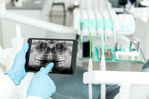
Digital X-Rays
Digital radiography (x-rays) is the latest technology used to take dental x-rays. This technique uses an electronic sensor (instead of x-ray film) that captures and stores the digital image on a computer. This image can be instantly viewed and enlarged on the computer screen. Digital x-rays reduce radiation by a large percentage compared to the already low exposure of traditional dental x-rays.
Dental x-rays are an essential diagnostic tool which provide vital information during a dental exam. We use this information to safely and accurately detect hidden problems and to complete a thorough comprehensive diagnosis. Without x-rays, problem areas may go undetected.
What an X-Ray can reveal
- Decay between the teeth
- Abscesses or cysts
- Bone loss
- Cancerous and non-cancerous tumours
- Developmental abnormalities
- Poor tooth and root positions
- Problems inside a tooth or below the gum line
Detecting and treating dental problems at an early stage may save you time, money, unnecessary discomfort, and more importantly your teeth.
Safety with dental radiography
Digital x-rays produce a significantly lower level of radiation compared to traditional dental x-rays. Not only are digital x-rays better for the safety of the patient, they are faster and more comfortable to take, which reduces your time in the dental practice. As the digital image is captured electronically, there is no need to develop the x-rays, thus eliminating the disposal of harmful waste and chemicals into the environment.
Even though digital x-rays produce a low level of radiation and are considered very safe, we still take necessary precautions to limit the patient’s exposure to radiation. These precautions include only taking x-rays that are necessary.
Frequency of dental x-rays
The need for dental x-rays depends on each patient’s individual dental health needs. Your dentist will recommend necessary x-rays based upon the review of your medical and dental history, a dental examination, signs and symptoms, your age, and risk of disease.
A series of dental x-rays is recommended for new patients. Bite-wing x-rays (x-rays of top and bottom teeth biting together) are recommended for most patients every 2 years.
For extractions, including wisdom teeth, an OPG (orthopantogram) is required to examine the tooth structure within the bone. When considering orthodontics an OPG and a lateral cephalogram are also needed.